Peritonsillar Space Infections
With the direct extension of tonsillitis, peritonsillar space infection occurs by exceeding the tonsillar capsule. It constitutes approximately 50% of deep neck infections(9). Aerobic and anaerobic agents cause infection. The most common causative agent is streptococcus pyogenes(10). Fever, sore throat, dysphagia or odynophagia, dehydration may be seen(11). A thickened and hoarse sound called 'hot potato' is typical in many patients (12). The patient cannot open his mouth fully and there is a trimus.
Diagnosis is physical examination, ultrasound, CT, and fine-needle aspiration to differentiate an abscess with cellulite. Treatment; In the presence of abscess, needle aspiration, incision and drainage, and warm tonsillectomy are in addition to IV antibiotic therapy(13). 90% improvement is achieved with needle aspiration, but more than one aspiration may be required. Incision and drainage may be required due to the high recurrence rate (14). Incision and drainage The incision is made where the anterior fold is most prominent and the abscess is drained with an oblique clamp. Warm tonsillectomy may be required in patients who extend into the lateral pharyngeal area and do not heal with drainage.
Submandibular Space Infections
In the past years; It was the most common site of DBE(15). Infections spread throughout the submandibular and sublingual area along the mylohyoid muscle in this region (16). 70-80% of the cases are of dental origin. Rigidity, trismus, sialorrhea, high fever and dysphagia are seen in the neck (17). If it reaches the parapharyngeal area, rapid airway obstruction may develop (18).
Ludwig's angina is a special infection. It is characterized by edema and faciitis of the facial planes without abscess (12). It involves the supramylohyoid and inframylohyoid areas. They spread through the neighborhood. Clinically, they spread aggressively and cause airway distress. Pain, dysphagia, salivary accumulation, neck rigidity, hyperemia in the floor of the mouth are seen.
Sublingual Area Infections
source of infection; teeth, lower lip, submandibular area and mandible. There may be a transition from the sublingual and submandibular areas to each other(19). The tongue is pulled inward due to induration and edema in the floor of the mouth. There is difficulty in swallowing.
Masticator Area Infections
This area; Like the submandibular area, it is the most common site of odontogenic infections (20). It is closely related to the parotid area, parapharyngeal area and submandibular area(21). There are clinical symptoms in the form of difficulty in swallowing, pain and swelling. Trismus may develop due to masseter and internal pterygoid muscle irritation.
Parapharyngeal Space Infections
Infections of this region usually occur with the spread of infections of the retropharyngeal space, masticator area, subandibular space, and peritosillar space 22). Pain, fever, dysphagia and trimus are seen. In connection with the structures in the parapharyngeal area; Horner's Syndrome, vena jugularis interna thrombosis and carotid rupture may develop. It can pass through the hazardous area or the carotid sheath into the mediastinum.
Retropharyngeal Space and Dangerous Cavity Infections
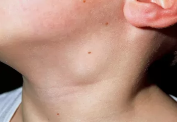
The most common cause in children is upper respiratory tract infections. In adults; esophageal instrumentation, trauma and vertebral fractures (Picture 2). It is more common in children, and torticollis in the neck is an important indicator (23). Infections of this region are the most dangerous because of the possibility of progression to the mediastinum and dangerous area. The dangerous area is located behind the retropharyngeal area. Between them is the alar fascia. In the dangerous area, it extends from the skull base to the diaphragm, and rapid infection can be transmitted to these areas (21). The patient has odynophagia, dysphagia, fever, sore throat and neck stiffness. The neck leans to one side. Breathing difficulties may occur. There is swelling on one side of the medial raphe in the oropharynx.
Parotid Area Infections
It occurs due to infection of parotitis or lymph nodes in the parotid. Due to its proximity to the parapharyngeal area, the infection can easily pass to this area.
Anterior Visceral Space Infections
It develops as a result of trauma to the hypopharynx or esophagus. It is risky because it can cause mediastinitis. Hoarseness and dyspnea may be the first signs. Dysphagia is seen.
Prevertebral Space Infections
Before the antibiotic era, cold abscess (Pott's abscess) due to tuberculosis was seen (24). Since there are vertebrae at the back, infections of this region occur as a result of penetrating injuries to the posterior pharyngeal wall or active spondylodiscitis.
The boundaries of the region are clear. Therefore, perforation of the fascia, spread to the dangerous area and mediastinitis are not common. In this area; Since there is no midline raphe, swelling is seen in the midline. Patient complaints are nonspecific. Neck pain that worsens with swallowing, dysphagia and, rarely, dyspnea may occur.
How is DBE Diagnosed?
Diagnosis has several steps. These; anamnesis and physical examination, laboratory tests and imaging methods.
- History and physical examination
Anamnesis and physical examination are the most important steps in diagnosis(25). The patient should be questioned about pain, dental problems, previous URTI, trauma, difficulty in swallowing, respiratory distress and diseases that impair immunity.
In addition to fever, weakness, loss of appetite, sore throat, swelling in the neck, dysphagia and trismus are quite common in patients (7).
Other symptoms depend on the space involved. Neck asymmetry due to neck mass or lymphadenopathy is seen in 70% of pediatric retropharyngeal abscesses. In parapharyngeal space involvement, the lateral pharyngeal band and tonsil become medialized. With the involvement of the paraspinal muscles, there is a decrease in torticollis and neck movements. Neural deficits may develop, especially in the cranial nerves. Leggy fever (as a result of internal jugular vein thrombophlebitis and septic embolization) may occur. Shortness of breath may occur due to airway obstruction and pulmonary complications.
-Laboratory Tests
In patients presenting with DBE; complete blood count, blood biochemistry tests and coagulation tests should be performed. Throat culture and blood culture may be required. If there is an abscess, the abscess material should be obtained during needle biopsy or drainage and sent to the laboratory for culture (21).
-Display Methods
Lateral neck radiographs: It is useful to see retropharyngeal abscesses, edema in the soft tissues of the prevertebral area, flattening of the cervical axis and gas formation in the soft tissue (26). The neck should be pulled in extension and during deep inspiration. Prevertebral and retropharyngeal soft tissue thickness is affected by position and breathing, as well as changes with crying and swallowing. If the thickness of this tissue is more than 7 mm at the C2 level (in the retropharyngeal region) and more than 14 mm at the C6 level (22 mm in adults) in children and adults, it can be interpreted in favor of infection.
Chest X-ray: It can show the presence of pneumonia or mediastinitis, as well as help detect lung pathologies such as tuberculosis.
Computed Tomography: It has the most important place in the diagnosis of deep neck infections and abscesses (27). While its sensitivity is 95%, it can go up to 100% if it is shot with contrast. Vascular structures, jugular vein thrombosis, lymph node anatomy can be revealed more easily with contrast material. It is useful in making a surgical indication (28). Classical findings of abscess on neck CT; low density in the abscess center, contrast enhancement around the mass, soft tissue swelling, mass effect and deletion of fatty planes (29).
DBE Treatment
Abscesses may progress with airway obstruction. The first step in treatment is airway control. Next is antibiotic therapy and drainage. Providing an airway is very important in all deep neck infections. Securing the airway is possible with intubation or tracheotomy. In patients with dyspnea and stridor, the airway should be checked and secured immediately.
Although it is an increasing view to treat patients with antibiotics alone, antibiotherapy is still used as an accompanying treatment method with surgery. Antibiotherapy should be given intravenously. It generally starts empirically. Antibiotherapy should be directed against the most common organisms, streptococci and anaerobes. The antibiotic chosen, especially in dental infections, should also be effective against anaerobic microorganisms (30). While penicillin was the first choice antibiotic in the past, the increase in beta-lactamase production led to a change in antibiotic choice (31). Antianaerobic agents should be used together with beta-lactamase-resistant penicillins. Clindamycin can be used if you are allergic to penicillin. Antibiotherapy should be arranged according to the culture result(12). Intravenous treatment is continued for 48 hours when the patient is fever-free. After that; Oral use can be started with amoxicillin-clavulanate, clindamycin or ciprofloxacin.
Surgical intervention is required in deeply located abscesses that obstruct the airway, if sepsis has developed, and in cases where there is no response despite 48 hours of antibiotic treatment. Surgical intervention should be planned according to the location of the abscess and its relationship with the structures in the neck. Peritonsillar and retropharyngeal abscesses are drained with a transoral approach. It is drained in the trendelenburg position due to the risk of aspiration in retropharyngeal abscesses. In the external approach, the incision site is determined according to the location of the abscess and drainage is provided.
DBE Complications
Complications of deep neck infections and the mortality rate due to these complications have decreased with the introduction of antibiotics (32). However, deep neck infections can be fatal if life-threatening complications develop(33). Death from deep neck infections; It may be due to airway obstruction, as well as after mediastinitis, (34) cavernous sinus thrombosis (35), pyothorax (36) and rupture of great vessels into the chest cavity (37). Rare complications such as cranial nerve paralysis, cervical necrotizing fasciitis, vena jugularis interna thrombosis, septic shock, DIC picture, renal failure, meningitis, epidural abscess can also be seen in DBE(38).
Conclusion
In conclusion; DBE are infections that develop in potential spaces surrounded by fasciae in the neck region. DBE is a serious clinical condition and may progress with death. When not recognized early or treated inadequately or inappropriately, they can lead to life-threatening complications as they can spread to adjacent spaces in the neck that contain vital structures.
References
1. Suehara AB, Gonçalves AJ, Alcadipani FA, Kavabata NK, Menezes MB. Deep neck infection: analysis of 80 cases. Braz J Autorhinolaryngol 2008;74(2):253-9.
2. Chen MK, When YS, Chang CC, Huang MT, Hsiao HC. Predisposing factors of lifethreatening deep neck infection: logistic regression analysis of 214 cases. J Otolaryngol 1998; 27(3):141-4.
3. Baker AS , Montgomery WW .Oropharyngeal space infections. Curr Clin Top Infect Dis 1987;8:227-65.
4. Goldstein NA, Hammerschlag MR. Peritonsillar, retropharyngeal, and parapharyngealabscess. In: Feigin RD, Demler GJ, Cherry JD, Kaplan SL, editors. Textbook of Pediatric Infectious Diseases. 5th ed. Philadelphia: WB Saunders: 2004, p:178-85.
5. Myers EM , Kirkland LS Jr , Mickey R. The head and neck sequelae of cervical intravenous drug abuse. Laryngoscope 1988;98(2):213-8.
6. Nusbaum AO , Som PM , Rothschild MA , Shugar JM . Recurrence of a deep neck infection: a clinical indication of an underlying congenital lesion. Arch Otolaryngol Head Neck Surg 1999;125(12):1379-82.
7. Marra S, Hotaling AJ. Deep neck infections. Am J Otolaryngol 1996;17:287-98.
8. Welsh L, Welsh J, Gregor F. Radiographic analysis of deep cervical abscesses. Ann Otol Rhinol Laryngol 1992;101:854-60.
9. Ungkanont K, Yellon RF, Weissman JL, Casselbrant ML, Gonzalea-Valdepena H, Bluestone CD. Head and neck space infections in infants and children. Otolaryngol Head Neck Surg 1995; 112: 375-82.
10. Steyer TE. Peritonsillar abscess: diagnosis and treatment. Am Fam Physician 2002;65(1):93-6.
11. Schraff S, McGinn JD, Derkay CS, Peritonsillar abscess in children: a 10 year review ofdiagnosis and management. Int J Pediatr Otorhinolaryngol 2001;57:213-8.
12. Weed HG,Forest L. Deep Neck Infections. Can Koç, Cummings Otolaryngology Head and Neck Surgery. Ankara: 4 editions. Sun Bookstore; 2007:2515-25.
13. Mr. İ. Deep neck infections. Clinical Development 2012;25:14-7.
14. Wolf M, Even-Chen I, Kronenberg J. Peritonsillar abscess: repeated needle aspirationversus incision and drainage. Ann Otol Rhinol Laryngol 1994;103:554-7.
15.Meher R, Jain A, Sabharwal A, Gupta B, Singh I, Agarwal AK. Deep neck abscess: a prospective study of 54 cases. J.Laryngol Otol 2005:119:299-302.
16. Ariji Y, Gotoh M, Kimura Y, Naitoh M, Kurita K, Natsume N, Ariji E. Odontogenicinfection pathway to the submandibular space: imaging assessment. Int J Oral Maxillofac Surg 2002;31:165-9.
17. Peterson LJ. Contemporary management of deep infections of the neck. J Oral Maxillofac Surg 1993:51:226-31.
18. Britt JC, Josephson GD, Gross CW. Ludwig's angina in the pediatric population: reportof a case and review of the literature. Int J Pediatr Otorhinolaryngol 2000:52:79-8.
19. Harnsberger HR. The oral cavity: emphasizing the sublingual and submandibular spaces.In: Harnsberger HR, ed: Handbook of Head and Neck Imaging. 2nd edn. St. Louis: MosbyYear Book, Inc. 1995: p120-49.
20. Ariji E, Moriguchi S, Kuroki T, Kanda S. Computed tomography of maxillofacialinfection. Dentomaxillofac Radiol 1991;20:147-51.
21. Dursun E, Eryılmaz E. Neck anatomy and infections. Can Koç, Otorhinolaryngology and Head and Neck Surgery. Ankara: 1 edition. Sun Bookstore; 2004:p773-875.
22. Harnsberger HR. The parapharyngeal space and the pharyngeal mucosal space. In:Harnsberger HR, ed: Handbook of Head and Neck Imaging. 2nd edn. St. Louis: Mosby YearBook, Inc.1995:p29-45.
23. Santos Gorjón P, Blanco Pérez P, Morales Martín AC, Del Pozo de Dios JC, Estévez Alonso S, Calle de la Cabanillas MI.Deep neck infection. Review of 286 cases. Acta Otorrinolaryngol Esp2012 ;63(1):31-41.
24. Hedge A, Mohan S, Lim WE. Infections of the deep neck spaces. Singapore Med J 2012;53(5):305-1
25.Tan PT, Chang LY, Huang YC, Chiu CH, Wang CR, Lin TY. Deep neck infections in children. J Micro Immune Infect 2001;34:287-92.
26. Barratt GE, Kopman CF Jr, Coulthard SW. Retropharyngeal abscess: a ten yearexperience. Laryngoscope 1984;94:455-63.
27. Holt RG, MacManus K, Newman NK, Potter JL, Tinsley P. Computedtomography in the diagnosis of deep infections. Arch Otolaryngol 1992; 108:693-6.
28. Har-El G, Aroesty JH, Shaha A, Lucente FE. Changing trends in deep neck abscess.Aretrospective study of 110 patients.Oral Surg Oral Med Oral Pathol 1994;77:446-50.
29. Vural C, Gungor A, Comerci S. Accuracy of computerized tomographyin deep neck infections in the pediatric population.Am J Otolaryngol 2003;24(3):143-8.
30. Huang TT, Tseng FY, Yeh TH, Hsu CJ, Chen YS. Factors affecting the bacteriology of deep neck infection: a retrospective study of 128 patients. Acta Otolaryngol.2006;126(4):396-401.
31. Parker GS, Tami TA. The management of peritonsilla abscesses in the 90s: an update.Am J Otolaryngol 1992;13:284-8.
32. Cottichia JM, Geoffrey S, Getnick GS, Yun RD, Arnold JE. Age-site and time specificdifference in pediatric deep neck abscesses. Arch Otolaryngol Head Neck Surg 2004;130:201-7.
33. Wang LF, Kuo WR, Lin CS, Lee KW, Huang KJ. Space infection of the head and neck. Kaohsiung J Med Sci 2002;18(8):386-92.
34. Pearse Jr. HE. Mediastinitis following cervical suppuration. Ann Surg 1938;108:588-611.
35. Feldman DP, Picerno NA, Porubsky ES. Cavernous sinus thrombosis complicatingodontogenic parapharyngeal space neck abscesses: a case report and discussion. OtolaryngolHead Neck Surg 2000;123:744-5.
36. Singh I, Chanda R, Gupta KB, Yadav SP. Fatal pyothorax: a rare complication ofretropharyngeal abscess. Indian J Chest Dis Allied Sci 2003;45:265-8.
37. Sallinger S, Pearlman SJ. Hemorrhage from pharyngeal and peritonsillar abscesses. Arch Otolaryngol 1933;18:464-509.
38. Lee JK, Kim HD, Lim SC. Predisposing factors of complicateddeep neck infection: an analysis of 158 cases. Yonsei Med J2007;48(1):55-62.