The neck is a region between the trunk and the head that allows the head to move quite freely. Its anatomy is quite complex due to the neural, vascular, respiratory and digestive structures it contains and can vary from person to person. The borders of the neck are formed superiorly by the lower edge of the mandible, temporal bone zygomatic process, external occipital protuberens, and inferiorly by the manubrium sterni, clavicle, acromioclavicular joint and the 7th cervical vertebra spinous process.
Neck Fascias(1,2)
The superficial layer of the deep fascia completely surrounds the neck and is therefore called the "Donating fascia" (investing fascia). It is also called the enveloping layer, the external layer, and the anterior layer of the deep fascia. This fascia is defined by the rule of two. Because it surrounds two muscles, two glands and creates two cavities. It originates from the spinous processes of the columna vertebralis and extends circularly in the neck, surrounding the sternocleideomastoid muscle and trapezius. It attaches to the hyoid bone in the midline and continues superiorly, packing the submandibular and parotid glands. Here it also surrounds the anterior abdomen of the digastric muscle and the mylohyoid muscle, thus forming the ceiling of the submandibular space. It fuses between the parotid and submandibular glands to form the stylomandibular ligament and attaches to the styloid process. It contributes to the formation of the lateral side of the carotid sheath. The fascia lateral to the parotid rises upward to form the temporal fascia. At the level of the mandible, the fascia divides into an internal layer, which surrounds the medial aspect of the medial pterygoid muscle up to the skull base. The outer layer surrounds the masseter and attaches to the zygomatic posterior. Inferiorly, it attaches to the clavicle, sternum, and acromion of the scapula. The two spaces formed by this fascia are the posterior triangular space on the same side and the “suprasternal space of Burns” in the midline. The suprasternal space arises when two layers of fascia attach to the front and back of the sternum. At the level of the mandible, the fascia divides into an internal layer, which surrounds the medial aspect of the medial pterygoid muscle up to the skull base. The outer layer surrounds the masseter and attaches to the zygomatic posterior. Inferiorly, it attaches to the clavicle, sternum, and acromion of the scapula. The two spaces formed by this fascia are the posterior triangular space on the same side and the “suprasternal space of Burns” in the midline. The suprasternal space arises when two layers of fascia attach to the front and back of the sternum. At the level of the mandible, the fascia divides into an internal layer, which surrounds the medial aspect of the medial pterygoid muscle up to the skull base. The outer layer surrounds the masseter and attaches to the zygomatic posterior. Inferiorly, it attaches to the clavicle, sternum, and acromion of the scapula. The two spaces formed by this fascia are the posterior triangular space on the same side and the “suprasternal space of Burns” in the midline. The suprasternal space arises when two layers of fascia attach to the front and back of the sternum.
The middle layer of the deep fascia is also referred to as the visceral fascia, prethyroid fascia, and pretracheal fascia. This layer is divided into two subsections; muscular part and visceral part. The muscular layer covers the infrahyoid strap muscles, the adventitia of the great vessels, while the visceral layer covers the pharynx, larynx, esophagus, trachea, and thyroid gland. The muscular layer attaches superiorly to the hyoid bone and thyroid cartilage, and inferiorly to the sternum and clavicle. The visceral layer runs downward into the superior mediastinum and continues with the fibrous pericardium, enveloping the thoracic trachea and esophagus. The visceral layer is attached to the hyoid bone and thyroid cartilage anterosuperiorly. Posteriorly, it wraps around the buxinator and pharyngeal constrictors and progresses towards the skull base. This part is also called “Buccopharyngeal fascia”. This section forms the anterior wall of the retropharyngeal space. Both layers contribute to the formation of the carotid sheath. This fascia connects posteriorly with the alar layer of the deep cervical fascia at the level of the first and second thoracic vertebrae. The fascia in front of the thyroid gland is called the "prethyroid fascia", and the fascia in front of the trachea is called the "pretracheal fascia". (Picture 1)
The deep layer of the deep fascia originates from the spinous processes of the cervical vertebrae and the Ligamentum nuchae. It is divided into two as “Anterior alar layer” and “Posterior prevertebral layer” at the level of transverse processes of cervical vertebrae. The alar fascia extends from the skull base to the second thoracic vertebra, where it joins with the visceral fascia. This fascia runs between the visceral leaf of the middle layer of the deep fascia and the prevertebral fascia. The prevertebral fascia runs immediately in front of the vertebral corpuscles and runs along the entire vertebra. Thus, it progresses from the base of the skull to the coccyx bone. It runs circularly around the neck and surrounds the vertebral muscles, the deep muscles of the posterior neck triangle, and the scalene muscle. This layer of fascia surrounds the brachial plexus and subclavian vessels. It then continues laterally as the axillary sheath. The area between the alar and prevertebral fascia is called the Danger Space.
The carotid sheath is a structure formed by all three layers of fascia and contains the carotid artery, internal jugular vein and vagus nerve. It starts at the skull base and runs in front of the anterior surface of the prevertebral fascia in the neck and enters the chest behind the clavicle.
Neck Muscles (2)
M. sternocleidomastoideus (SCM): It begins by attaching to the apex of the mastoid bone above, divides into the sternal and clavicular bundles as it descends, and ends by attaching to the sternum and clavicle. It is surrounded by the superficial layer of the deep cervical fascia. From the surface of the muscle v. passes into the jugularis externa. The motor innervation of the muscle n. provides accessories.
Platysma: This thin muscle located in the anterior part of the neck is surrounded by the superficial neck fascia. It begins by attaching to the lower edge of the mandible corpus above, the skin of the lower part of the face and the muscles around the mouth commissure, and as it descends, it ends in the superficial fascia near the clavicle. It provides tension of the neck skin, at the same time it pulls the lower lip, mouth commissure and mandible slightly down. Motor innervation n. provides the cervical branch of the fascia.
M. digastricus: It is a two-bellied muscle, anterior and posterior. The anterior larynx begins by attaching to the posterior surface of the mandible corpus and proceeds in a band to attach to the greater horn of the hyoid bone. The posterior belly begins by attaching to the apex of the mastoid bone, progresses anteriorly and downwards to join with the anterior abdomen. The tendon at the junction of both abdomens is called the digastric tendon. Anterior belly of the muscle, n. Photos of mandibularis in Instagram Account n. branch of mylohyoideus, posterior belly n. It is innervated by the digastric branch of the fascia. The muscle's job is to lift the hyoid bone and pull the mandible down.
M. stylohyoideus: It begins by attaching to the styloid process, goes downwards and ends by attaching to the greater horn of the hyoid bone. It is innervated by the stylohyoid branch of N. facialis.
M. mylohyoideus: It begins by attaching to the linea mylohyoidea on the inner surface of the mandible corpus, coming towards the midline and anteriorly to the opposite m. fuses with mylohyoideus. It is attached posteriorly to the hyoid bone. The muscle elevates the floor of the mouth during swallowing, allowing the tongue to attach to the palate. motor innervation n. provides the mylohyoid branch of the mandi aris.
M. geniohyoideus: It begins by attaching to the inner surface of the mandible corpus, progresses backwards and attaches to the corpus of the hyoid bone. motor innervation. n. supplied by the hypoglossus. When it contracts, it lifts the hyoid bone and larynx and aids in the swallowing process.
M. sternohyoideus: It begins by attaching to the manibrium sterni and clavicle head below, and ends by attaching to the hyoid bone corpus above. Motor innervation is provided by the ansa cervicalis.
M. omohyoideus: It is a double-bellied muscle, upper and lower. Its upper abdomen is attached to the corpus of the hyoid bone, and its lower abdomen is attached to the scapular cleft of the scapula. The upper abdomen goes down and outward, and the lower abdomen goes up and inward and joins with a tendon. The tendon is attached to the clavicle and first rib by a fibrous band formed by the deep cervical fascia. Motor innervation is provided by the ansa cervicalis. Its function is to pull the larynx and hyoid bone down.
M. sternothyroideus: Its lower end is attached to the manibrium sternie and its upper end is attached to the outer surface of the thyroid cartilage. Motor innervation is provided by the ansa cervicalis. Its job is to pull the larynx down.
M. thyrohyoideus: Its upper end is attached to the hyoid bone corpus, and its lower end is attached to the outer surface of the thyroid cartilage. Its motor innervation is provided by the ansa cervicalis. Its job is to pull the larynx down.
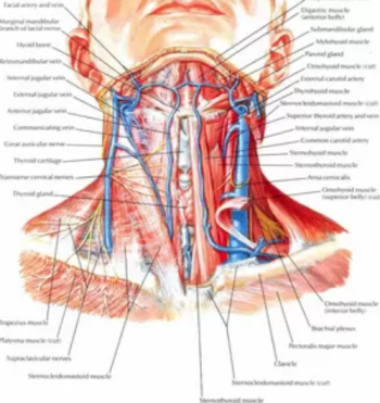
Neck Arteries (2,3)
1 A. subclavia and its branches (most importantly a. vertebralis)
2 A. carotid communis
A. carotid interna (does not give branches in the neck)
A. carotid externa and its branches
A. Carotis Communis:
Right, at the level of the sternoclavicular joint a. It separates directly from the aortic arch in the upper mediastinum on the left from the brachiocephalus. It progresses upward in the carotid sheath and divides into internal (AKI) and external (AKE) branches at the level of the upper edge of the thyroid cartilage, in the area called the bifurcation.
A. Carotid interna : It goes from the bifurcation to the skull base without branching. From the outer surface n hypogossus, a. occipitalis and m. The posterior part of the digastricus crosses the artery. AKI's v. jugularis interna (VJI), IX., X., XI. and XII. It has important relations with cranial nerves and ACE. IX., X. and XI. cranial nerves are between these two vessels. Further down, the VJI is located outside the AK1.
A. Carotid externa: The ACC courses outside the carotid sheath. After leaving the bifurcation m. passes under the posterior abdomen of the digastricus, m. styloglossus and m. It crosses the stylopharyngeus muscles superficially, enters the parotid gland, where it divides into terminal branches behind the condylar process of the mandible.
1. A. thyroidea superior: A. is the first anterior branch of the carotid externa, and it supplies the upper part of the thyroid gland along with some parts of the larynx and SCM muscle. It separates just below the cornu majus of the hyoid bone.
2. A. pharyngea ascendens: It is the smallest branch of A. carotid externa. It supplies the pharynx, soft palate, tonsil, middle ear and part of the meninges.
3. A. lingualis: It separates just above the A. thyroidea superior. n. The hypoglussus is located on the outside of the artery. artery m. It reaches the tongue by passing under the hyoglossus. a.lingualis; rear edge m. posterior abdomen of digastricus, anterior margin of m. anterior abdomen of digastricus, upper edge of n. The "Lesser triangle" determined by the hypoglossus can be connected with the way of arrival. Artery, m in the posterior part of the triangle. It can be found just deep to the hyoglossus.
4. A. fascialis: It is separated from the anterior surface of the external carotid. It progresses anteriorly and upwards from the depth of the M. digastricus. It is closely related to the submandibular gland. The artery, which deepens from the lower part of the gland, emerges from the upper level of the gland and becomes superficial. Crossing the lower edge of the mandible, the branches in the neck a. palatine ascendens, a. tonsillaris, submandibular gland branches and a. gives submentalis.
5. A. occipitalis: It arises from the posterior aspect of the external carotid at the level of separation of the facial artery. The artery supplies the SCM muscle, digastric and stylohyoideus muscles, and the suboccipital region of the scalp.
6. A. auricularis posterior: It is separated from the posterior face at the level of the upper edge of the digastric muscle. The parotid gland gives branches to the auricle and scalp.
7. A. temporalis superficialis: It is the terminal branch of the carotid externa.
8. A. maxillaris: It is the terminal branch of the carotid externa.
Neck Veins (2,3)
V. jugularis interna (VJI): Carotid artery and n. It is located in the carotid sheath with the vagus. It starts from the jugular forarne at the skull base, extends to the thoracic inlet, and v. brachiocephalic and v. terminates at the junction of the subclavian.
V. jugularis externa (VJE): It is located on the outer surface of M. SCM. Starting from the heme of the parotid tail, it extends downwards and extends to m in the posterior triangle. anterior to scalenius anterior v. ends in the subclavian. Posterior branch of V. retromandibularis and v. arises from the fusion of the auricularis posterior.
V. jugularis anterior (VJA): It provides drainage of the anterior part of the neck. It usually starts as a resultant vein from the submandibular region and progresses from the outside towards the midline on the anterior surface of the neck from top to bottom. Both VJA are connected by the jugular arch just above the sternum. Below the arc, the VJA is curved outward and either the VJI or the v. drains into the subclavian.
V. fascialis: Passing through the lower edge of the mandible, posteriorly v. It merges with the anterior branch of the retromandibularis. Behind this junction, the vein is called the common facial vein. Yen a. lingualis, n. The hypoglossus joins the VJI by proceeding from the outside of the AKI and ACE.
V. lingualis: Although the lingual veins are variable, they usually follow two paths. The dorsal lingual vein takes the drainage of the dorsum of the tongue, together with the a.lingualis m. Passing deep into the hyoglossus, it joins the VJI by the greater horn of the hyoid bone. The deep lingual vein can be seen under the mucous membrane on the underside of the tongue. The vein formed by merging with V. sublingualis, m. on the hyoglossus muscle n. It is closely related to the hypoglossus. Then either v. joins the lingualis or directly to the VJI.
V thyroidea superior: It is closely adjacent to A. thyroidea superior. Not only the thyroid gland, but also the larynx-related v. laryngeus superior and v. It also receives venous drainage of the cricothyroideus. v. Inferior thyroidea: Located anterior to the trachea and drains blood from the Isthmus of the thyroid gland into the left brachiocephalic vein located behind the manibrium of the sternum.
V. retromandibularis: Accompanying the two terminal branches of the external carotid artery, v. temporalis superficialis and v. The maxillary vein joins together in the parotid gland to form the retromandibular vein. Angulus mandible v. retromandibularis divides into anterior and posterior branches.
V. brachiocephalicus: VJE merges with VJI at the root of the neck and forms v. forming the brachiocephalicus v. It drains into the subclavia. Both v. The brachiocephalicus fuses to form the superior and na cava.
Neck nerves (2,3)
N. Glossopharyngeus: 9th nerve, n. The glossopharyngeus enters the neck by passing through the foramen jugulare together with the 10th (N. Vagus) and 11th (N. Accessorius) cranial nerves. branches n. tympanicus (sensory), lesser petrosal nerve (parasympathetic), carotid branch or branches (sensory), pharyngeal branches (sensory), tonsillar branches (sensory), lingual branches (sensory), and n. stylopharyngeus (motor).
Nervus vagus: It is the 10th cranial nerve and has sensory, motor and parasympathetic fibers. It arises from the brain stem, via the foramen jugulare v. jugularis interna and a. It continues its downward path between the carotis.
The neck branches of N. vagus are as follows;
Meningeal branches (sensory)
N. auricularis (sensory)
Pharyngeal branch (motor): It forms the pharyngeal plexus with branches from the N. glossopharyngeus and sympathetic trunk. It is the main motor nerve for the muscles of the pharynx and palate.
N. laryngeus superior: It starts from the inferior ganglion of the vagus. It reaches the larynx from the depths of the AKi and AKE. It divides into internal and external branches. The internal branch passes between the middle and lower constrictor muscles and provides sensation to the larynx. The external branch is under the lower constrictor muscle a. passes together with the thyroidea superior and m. It innervates the cricothyroideus.
N. laryngeus iriferior (recurrent laryngeal nerve-RLS): The right RLN starts from the lower part of the neck. N. vagus a. It leaves just anterior to the subclavian, curves downward, enters the larynx through the tracheoesophageal groove upwards behind the artery. Left RLS thorax n. It begins where the vagus crosses the aortic arch. RLSs pass under the lower border of the inferior constrictor muscle of the pharynx and cover the mucosa of the larynx and m. They innervate all intrinsic muscles except the cricothyroideus.
Nervus accessorius: 11th cranial nerve. As soon as it exits the foramen jugulare, it is directed towards the posterior triangle of the neck. It usually crosses the VJi externally as well as internally. M. It enters the muscle from the upper part of the SCM, after it moves through the muscle, it enters the posterior triangle of the neck. M. trapezius, before entering m in the posterior triangle. The levator runs over the scapula.
Nervus hypoglossus: It is the 12th cranial nerve. It leaves the cranial cavity from the hypoglossal canal of the occipital bone and reaches deep into the carotid sheath. It runs downwards under the posterior abdomen of M. digastricus, and v. jugularis interna and a. It comes out through the carotid interna. After turning around A. occipitalis, it moves forward to m. It proceeds deep into the submandibular gland above the hyoglossus muscle and distributes to the tongue muscles.
Ansa cervicalis: The strap in front of the larynx provides innervation to the muscles. Ansa cervicalis consists of lower and upper branches. Upper branch called ansa hypoglossia, n. hypoglossus a. It is the branch that the occipitalis gives as it turns around. The lower branches consist of C2-C3 fibers separated from the plexus cervicalis.
Cervical Plexus: Cervical plexus is formed by Cl-C4. It includes sensory, motor, and sympathetic branches.
N. occipitalis minor (Lesser occipital nerve)
N. auricularis magnus (Greater auricular nerve)
N. transverse colli (Transverse cervical nerve)
N. supraclavicularis
N. phrenicus: It is the motor nerve of the cervical plexus. Most of its stimuli come from C3 and C4, and a small part from C5. The phrenic nerve runs downward on the anterior scalene muscle, under the prevertebral fascia. It enters the thorax deeply through the subclavian vein and over the subclavian artery.
Cervical Lymph Groups (4,5)
Zone I: Includes submental and submandibular lymph node groups. The submental nodes are between the two anterior bellies of the hyoid bone and digastric muscle. The submandibular group is bounded by the mandible, anterior and posterior bellies of the digastric muscle. Level I lymph nodes are divided into submental (Zone la) and submandibular (Zone Ib) lymph nodes .
Zone II: Also known as upper deep jugular (jugulo-digastric) lymph nodes. They are adjacent to the upper third of the internal jugular vein between the skull base and the hyoid bone or carotid bifurcation. Anteroposteriorly of the 11th cranial nerve is divided into Region Ila, and those above posteriorly are divided into Region IIb.
Zone III: Medium deep jugular lymph nodes. They are between the hyoid bone above and the omohyoid muscle below.
Region- IV: It is the lower deep jugular lymph nodes. The upper border is the omohyoid muscle, the lower border is the clavicle. These are the lower deep jugular lymph nodes.
Region V: It includes all lymphatic groups in the posterior triangle between the posterior border of the SCM muscle, anterior trapezius muscle, and the clavicle. There are three main lymphatic groups in the posterior triangle. 11 Lymph nodes around the 1st cranial nerve, lymph nodes along the transverse cervical artery, and supraclavicular lymph nodes form these three groups.
The posterior triangular lymph nodes are divided into Zone Va, those above the posterior abdomen of the omohyoid muscle, and Zone Vb, those below.
Zone VI: Includes lymph nodes in the anterior neck compartment. They are located in the midline between the hyoid bone and the suprasternal notch. On both sides, the carotid arteries form the lateral borders. The anterior compartment (prelaryngeal, paralaryngeal, pretracheal, paratracheal, perithyroidal and Delphian) contains lymph nodes.
Region VII : Lymph nodes in the anterosuperior mediastinum and tracheoesophageal groove. (Picture 2)
K bib