The larynx is a part of the respiratory tract and is located in the anterior middle part of the neck, under the chin, between the pharynx and the trachea. It is a mobile organ, it moves up and down with swallowing.
The top of the larynx consists of a pyramid-like anatomical structure below, cartilage, muscles, and connective tissues. The larynx consists of 3 parts from top to bottom.
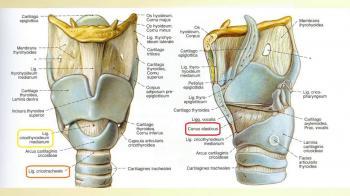
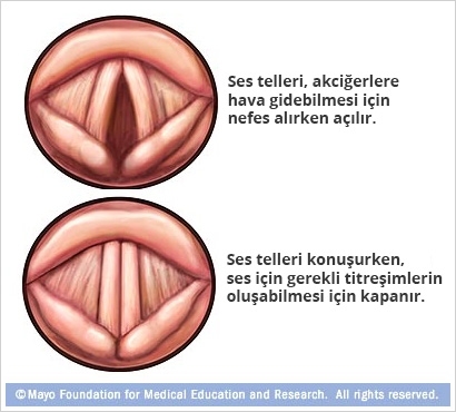
The upper part of the vocal cords called supraglottic at the top, the vocal cords section that forms the rare place of the larynx in the middle, the glottic section, and the vocal subglottic section below. Cartilage structures form the roof of the larynx with cartilage. There are 3 single cartilages and 3 bilateral cartilages.
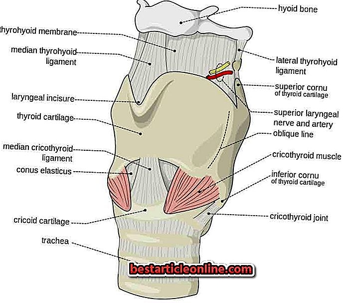
The epiglottis is a leaf-shaped cartilage that expands upwards, with a single cartilage. During swallowing, it closes the larynx and prevents food from escaping into the trachea. Thyroid Cartilage (Adam's apple-Cartilago thyroidea) It is a large cartilage in the form of a book leaf with an opening facing the back. Vocal cords, false vocal cords and muscles, which are important in voice formation, attach to this cartilage in the anterior part. The cricoid cartilage (cartilago cricoidea) is in the form of a thimble and is located anteriorly between the wider trachea and the thyroid cartilage.
Double Cartilage Aritenoid articulates with the posterior upper part of the cricoid cartilage. It is the attachment site of the posterior parts of the vocal cords. There are two supporting cartilages attached to the upper and lateral parts of the arytenoid. These are the corniculate and cuneiform cartilages. Laryngeal Ligaments (Ligeman) The skeletal roof of the larynx is connected to the neck by elastic structures from above and below. Main upper carrier ligament Temporol Bone (Ear)'a hyoidbetween the bones. The opening of the hyoid bone is a large bone that faces posteriorly and provides the movement of the larynx with various ligaments and muscles.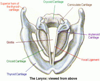
Thyrothyroidoid membrane; This membrane has a fibroelastic structure and is in the form of a wide curtain between the hyoid bone and the thyroid cartilage.
Quadrangular membrane: It is a wide, quadrangular membrane located between the arytenoid cartilage and the epiglottis cartilage.
Conus elasticus membrane: (Cricovocal membrane) It is a curtain that starts from the cricoid cartilage and continues towards the vocal cords and attaches to the vocal protrusion of the arytenoid cartilage from the thyroid cartilage, which we call the vocal ligament.
Vocal ligament: the upper free edge of the conus elesticus thickens and a taut ligament is formed along the free edge of the vocal cord by attaching the thyroid cartilage anteriorly to the vocal prominence posteriorly, this is what we call the vocal ligament. Here, together with the vocal ligament, the epithelial layer that covers it forms the vocal cords.
internal and external (extrinsic, intrinsic) is divided into two groups. Extrinsic (External) muscles provide movement and fixation direction of the larynx as a whole; muscles that lift the larynx up functionally (Digastric, stylohyoid, geniohyoid, mylohyoid muscles.) Depressor muscles that lower the larynx (thyrohyoid, sternohyoid, amohyoid and sternothyroid muscles.)
These muscles in the larynx structure control the airway. They control the resistance of the air between expiration (breathing) during phonosion (during sound production), and also prevent foreign substances from escaping into the lower respiratory tract during swallowing.
The cricothyroid muscle is actuated by the outer branch of the superior laryngeal nerve and the other muscles by the inferior laryngeal nerve. All these muscles are bilateral except the arytenoid muscle.
Cricothyroid muscle: this muscle has a significant influence on the position of the vocal cords (vocal cords). Its function in phonation (sound production center) is to increase the tension of the vocal cords. The muscles that provide tension of the vocal cords are the cricothyroid and thyroarytenoid muscles. The cricothyroid muscle is between the cricoid and thyroid cartilages. The thyroarytenoid runs parallel to the vocal ligament and under strong stresses the jack works against the thyroid.
The thyroarytenoid muscle stabilizes the position of the two cartilages, stretches the vocal cords and increases their length.
The cricothyroid muscle is usually relaxed at low frequencies. However, it contracts strongly in falsetto or high tone. Thanks to its synergistic (harmonious) work with the posterior cricoarytenoid muscle, the vocal cord space is maximally enlarged.
The cricothyroid muscle plays an important role in the control of foundational frequency in phonation and respiration. Arytenoid muscle: It is a single muscle located on the posterior surface of both arytenoid cartilages. It has straight and cross fibers. This muscle brings the arythyroids closer together and provides the approach (adduction) of the vocal cords.
Cross fibers narrow the entrance to the larynx, while straight fibers narrow the vocal space of the cord (sphines).
Thyroarytenoid muscle: It is useful to examine it as two separate parts. 1st part inner musculus (musculus vocalis)
Vocal muscle bundle musculus tatirol thyrooritioid exsternus (external) musculus vocalis (voice muscle or internal thyroarithyroid muscle) Anteriorly, the laryngeal surface of the thyroid cartilage arises from the middle part and attaches to the free edge of the concelasticus and lateral to the vocal ridge. The vocal muscle is attached to the vocal ligament. They are located in the vibrating part of the vocal cord.
The musculustriaritoid exsternus (Outer bundle) attaches from the middle part of the laryngeal surface of the thyroid cartilage posteriorly between the vocal process and the lateral (lateral) cricoarytenoid. Unlike the inner bundle, the cord extends along the outer lateral neighborhood of the vocalon and attaches to the arytenoid cartilage.
Thyroarytenoid Muscle Rotates the arytenoid cartilage anteriorly and inward. It is the opposite of the cricothyroid and posterior urcrico arytenoid muscle. It provides the task of closing the false vocal cord by bringing it closer to each other (sifintel) together with the transfers arithyroid muscle. This muscle brings the vocal cords closer together, stretches them and increases their thickness. It is the most important muscle for phonation. (It is the muscle of the vocal cord itself.) Therefore, the thyroaryroid muscle contains two bundles. The Outer Bundle contains fast-twitch fibers and provides dynamic movements. The vocal part (inner bundle) controls the backbone of the vocal fold. The outer bundle (Cotiral compartment) provides the closure of the vocal cords. Approximation of the vocal muscle (inner bundle) can be divided into two as inferule and spherule (lower and upper).
It has been suggested that the lower part provides the tension of the vocal cord at the supplogative level (the lower face of the vocal cord), the phendomental frequencies of the end of the vocal cord vibration control the phonation, and the upper part ensures the sound quality and especially the formant frequencies.
The posterior cricoarytenoid muscle attaches to the pits on the sides of the midline on the posterior surface of the croleoid cartilage, and its fibers extend outward and to the tip, extending to the lateral projection surface of the arytinoid. With the movement of this muscle, the arytinoid cartilages turn sideways and backwards, and as a result of this rotation movement, the vocal cords move away from each other. This is called the Abdulesion movement. This muscle is the main abductor of the vocal cord. (Opening muscle) during phonation acts opposite to the stretching function of thyroarytinoid and jack thyroid muscle and relaxes it.)
The lateral cricoarytenoid muscle is attached to the anterior surface of the lateral (outer protrusion) projection of the arytinoid, starting from the Dos-upper surface of the cricoid arch (arch) and the conus elastus and moving upwards posteriorly. This small quadrangular muscle pulls the arythyroid cartilage anteriorly and outward, bringing the vocal cords closer to the middle. (Adductor Function) The posterior crico is the opposite aspect of the arythroid muscle.
In terms of function, the vocal cords are divided into 3 histological divisions.
1. Cover a) Epithelium
b) The lucial layer of the Lamina Propria.
2. Passage a) Middle layer of laminol propria
b) Deep layer
3. Trunk Vocal muscle fibers